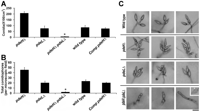
Figure 2. PdeH function regulates asexual development in M. oryzae.
Comparative quantitative analysis of conidiation (A) and conidiophores (B) in the wild type, pdeHΔ, pdeLΔ, pdeHΔ pdeLΔ and the complemented pdeHΔ strain (Comp pdeHΔ; RFP-PdeH expressing pdeHΔ). (A) For the assessment of conidiation, the respective strains were initially grown in the dark for a day followed by exposure to constant illumination for 6 d. Data represents mean ± SE based on five independent replicates. (B) The number of conidiophores produced by the indicated strains per microscopic field was quantified 24 h post photo induction. Results were quantified in three independent replicates and represented as mean ± SE. Asterisk highlights the heavily pigmented aberrant structures of varied morphologies produced by the double deletion mutant during conidiation phase. The quantification represents conidia-like or aberrant conidiophore like structures present therein. (C) Total number and arrangement of conidia on conidiophores in the wild type, pdeHΔ, pdeLΔ and the pdeHΔ pdeLΔ. The indicated strains were grown on PA medium layered on a glass slide. The strains were initially incubated in the dark for a day and then subjected to constant illumination for a period of 3 d prior to microscopic observations. Inset depicts the rare two celled conidia-like structure formed by the double deletion mutant. The arrow points to the single septum in the aberrant structure. Scale bar = 25 micron.

