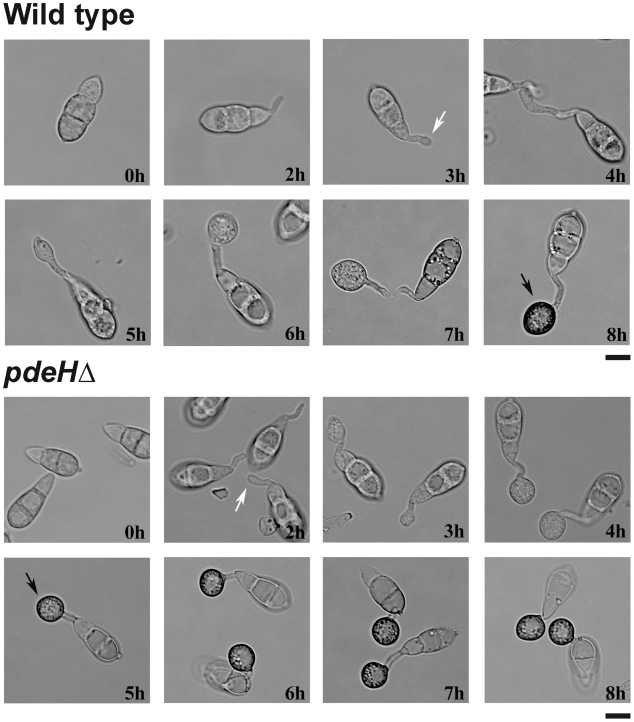
Figure 5. Loss of PdeH function advances appressorium formation and maturity.
Comparative time-lapse observation of germination and appressorium formation in the wild-type or pdeHΔ conidia. Equivalent number of conidia from the indicated strains were inoculated on cover slips and incubated in a moist chamber at room temperature. The samples were analysed and micrographed every hour over an 8 h period. White arrows indicate the stage of appressorium initiation/germ tube hooking, whereas black arrows depict melanized appressoria. Scale bar = 10 micron.

