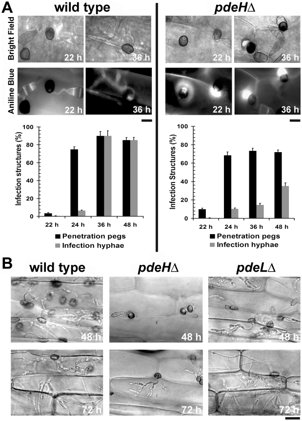
Figure 7. The pdeHΔ mutant is defective in its ability to colonize the host tissue.
Analysis and quantification of host penetration and in planta development in the wild type and pdeHΔ. (A) Photomicrographs depicting aniline-blue stained host papillary callose deposits in the wild type or pdeHΔ at the specified time points post inoculation. Scale bar = 10 micron. Bar charts representing effective host penetration (black bars) as well as the efficiency of the subsequent development of infection hyphae (gray bars) in the indicated strains. (B) Microscopic observations tracking the development of infection hyphae and the ability to spread and colonize the host tissue. Micrographs depict in planta growth in the indicated strains, 48 and 72 hpi on barley leaf explants. Prior to microscopic observations, the inoculated leaf samples were clarified using methanol and stained with acid fuchsin. Scale bar = 25 micron.

