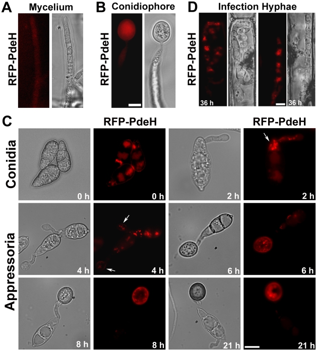
Figure 10. Subcellular distribution of RFP-PdeH fusion protein during different stages of pathogenic and asexual development.
(A) Vegetative hyphae from RFP-PdeH strain were imaged after 3 d growth on PA medium. (B) Mycelial blocks of the RFP-PdeH strain were exposed to constant illumination on fresh agar medium, and developing aerial (conidiophore) structures imaged after 24 h. Scale Bar = 10 micron. (C) Conidia harvested from the RFP-PdeH strain were inoculated on plastic cover slips and incubated in a moist chamber prior to microscopic observations. Bright field and epifluorescence images were captured at the indicated time points, using the requisite filter sets. The arrows highlight the localization pattern of RFP-PdeH fusion protein at 2 hpi (punctate) and 4 hpi (plasma membrane). Scale Bar = 10 micron. (D) RFP-PdeH conidia were inoculated on rice leaf sheath and the infection hyphae imaged after 36 h using epifluorescence microscopy. Scale Bar = 10 micron.

