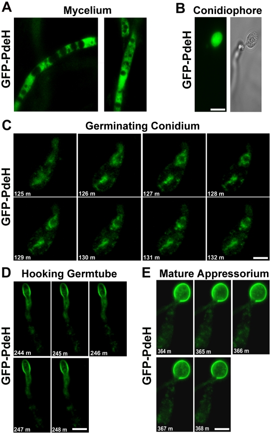
Figure 11. Localization and dynamic nature of the PROMpg1-GFP-PdeH during various stages of development in M. oryzae.
(A) The PROMpg1-GFP-PdeH localized to the cytosol in the vegetative hyphae and (B) developing aerial structures (conidiophore). Scale Bar = 10 micron. (C), (D) and (E) Snapshots extracted from time-lapse movies (supplemental movies), showing the highly dynamic PROMpg1-GFP-PdeH, during different stages (specified sequentially) of pathogenic development. Scale Bar = 10 micron.

