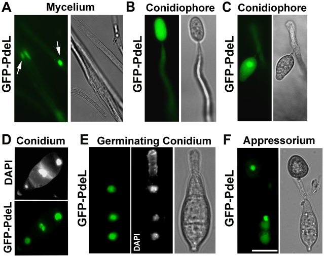
Figure 12. Predominant nuclear localization of the PROMpg1-GFP-PdeL during asexual and pathogenic development in M. oryzae.
(A) Vegetative mycelia from a PROMpg1-GFP-PdeL colony, showing the nuclear distribution of GFP-PdeL (arrows). (B) and (C) GFP-PdeL in aerial structures such as an immature and a mature conidiophore respectively. (D) Conidia from PROMpg1-GFP-PdeL strain were stained with DAPI and assessed using epifluorescence microscopy. (E) A germinating conidium incubated on an inductive surface for 2 h, and stained with DAPI while (F) shows an epifluorescence microscopic image of GFP-PdeL in a mature appressorium formed after 12 h incubation on a plastic coverslip. Bright field images outline the fungal structures in each instance. Scale Bar = 10 micron.

