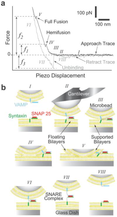
Fig. 1.

AFM force vs. piezo displacement measurement of interactions between opposing lipid bilayers and SNAREs. (a) A typical AFM force scan measurement showing hemifusion and full fusion of the compressed lipid bilayers and the unbinding of the SNARE complex during approach and retraction of the cantilever, respectively.21 The compression force required to induce bilayer hemifusion (f1) and fusion (f2) is measured at the onset of each event. Here, we generically refer to f1 as the fusion force. Alternatively, the unbinding force (f3) is measured during the sharp transition in the retraction trace as the SNARE complex dissociates under pulling. (b) Cartoon of our experimental system (not to scale) depicting the different steps (roman numerals) during the force scan measurement shown in (a). Lipid bilayers were formed on the glass dish and glass microbead attached to the cantilever tip. Upon approach of the cantilever toward the substrate, SNAREs embedded in the opposing bilayers form a complex (II) and the bilayers hemifuse (IV) and fully fuse (V) under compression. During the retraction phase, the SNARE complex is extended (VII) before it dissociates (VIII) under pulling.
