
Figure 1.
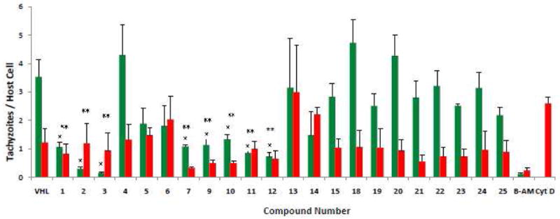
Quantification of invasion/attachment inhibition using red/green assay. The red/green invasion assay was performed using published methods.24 Tachyzoites (5 × 10⁶) were mixed with DMSO [vehicle (VHL)] or compound (final 10 μM) and allowed to sit at room temperature for 20 min. before being added to HFF monolayers growing in 8-well chamber slides. After 1 hour at 37 °C, 5% CO2, the cells were rinsed and fixed. Attached/extracellular parasites were detected using Rb anti-p30 (SAG1) (AbD Serotec, UK) followed by Alexa Fluoro 594 (red) (Invitrogen, CA). After permeabilization, penetrated/intracellular parasites were stained with MAb 9e11 anti-SAG1 (Argene Inc., NY) followed by Gt antimouse Alexa Fluoro 488 (green). DAPI (Invitrogen) for staining nuclei was added to secondary antibody. BAPTA-AM (20 μM) and cytochalasin D (2 μM) are included as controls for defects in attachment and penetration, respectively.24 Stained cells were examined by phase contrast and reflected fluorescence using an Olympus BX41 microscope. Numbers of green and red tachyzoites per host cell were enumerated by visual counting. Data are mean values ±SEM of three independent experiments, counting 10 random fields per well at 600× magnification. A single asterisk indicates tachyzoite penetration significantly lower (P ≤ 0.05, two-tailed Students' t-test) than vehicle. A double asterisk indicates a significant effect (P ≤ 0.05, one-tailed Students' t-test) on parasite attachment relative to vehicle.