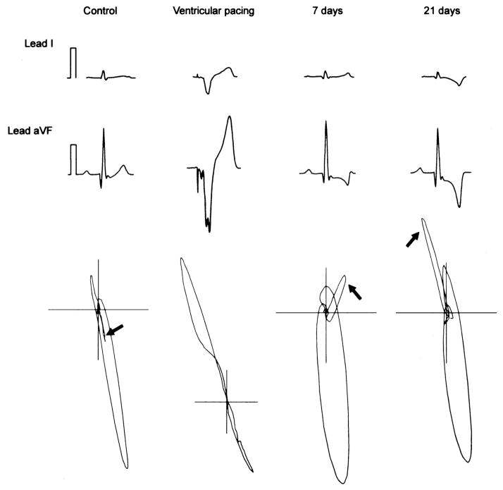
Figure 1. Time course of cardiac memory.
Surface ECG leads I and avF from a canine model of ventricular pacing to induce memory is illustrated. ECG’s and VCG’s recorded before onset of pacing, during pacing and at 7, 21 days after ventricular pacing are illustrated. The arrows indicate the change in T-wave vector which follows the vector of the paced QRS indicative of memory (adapted from Yu et al.)

