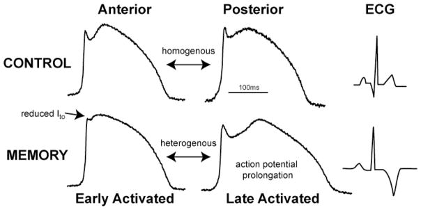
Figure 2. Action potential remodeling in cardiac memory.
The top panel illustrates action potentials recorded using optical imaging from unpaced dogs. The action potentials from anterior and posterior walls are similar with minimal regional action potential gradients. In contrast, following memory there is marked action potential prolongation in late-activated region. Also note the significant attenuation of epicardial phase1 notch limited to the early-activated region (arrow). This heterogeneous action potential remodeling causes regional repolarization gradients which underlies the electrophysiological basis for T-wave memory.

