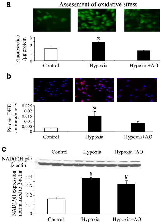
Fig. 3.
Assessment of oxidative stress. a Top Representative fluorescence staining of the oxidative stress conversion of 2′,7′-dichlorodihydrofluorescein diacetate (DCHFDA). Bottom Fluorescence quantification of the conversion of DCF (expressed as fluorescence per microgram protein). b Top Representative fluorescence staining of the oxidative stress conversion of dihydroethidium (DHE). Bottom Quantification of the percent area where DHE staining was present, normalized by the number of nuclei in each microscopic field analyzed. c Protein expression bands and densitometric analysis of NAD(P)H oxidase. Under hypoxic conditions, there was increased protein expression of NAD(P)H p47phox, together with an increase in the amount of reactive oxygen species suggesting a pro-oxidant state. In summary, hypoxia led to a pro-oxidant state that was improved when cells were treated with antioxidants, mainly through the metabolism of the increased reactive oxygen species. *p<0.05 compared to control and hypoxia + AO, ¥p<0.05 compared to control. These data (Figs. 2 and 3) suggest that increased oxidative stress plays a role in the decreased stem cell viability under hypoxic conditions.

