Fig. 5.
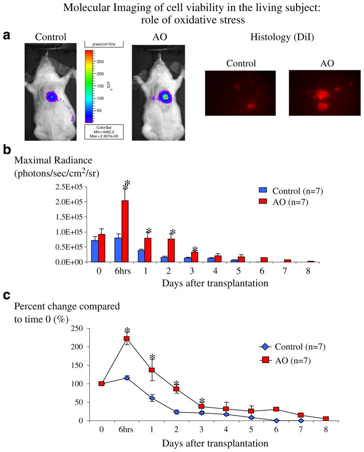
Noninvasive monitoring of cell engraftment and survival in living rats (n=7 in each group). a Representative images from control and AO animals 6 h after cell transplantation (left) and histologic confirmation (using the cell marker DiI, right) of the increased survival observed in AO animals compared to controls, showing the increase in early stem cell engraftment observed in AO animals, compared to controls. b, c Longitudinal monitoring and quantification of cell viability in living subjects. Data is expressed as maximal radiance (photons/sec/cm2/steridian, b) and as percent change (%) of maximal radiance compared to day 0 (c). Until day 3, cells that were treated with antioxidants (AO) had higher myocardial engraftment and survival compared to untreated cells (control). After day 3, cell survival was similar between the two groups. Error bars represent SEM. *p<0.05 compared to day 0.

