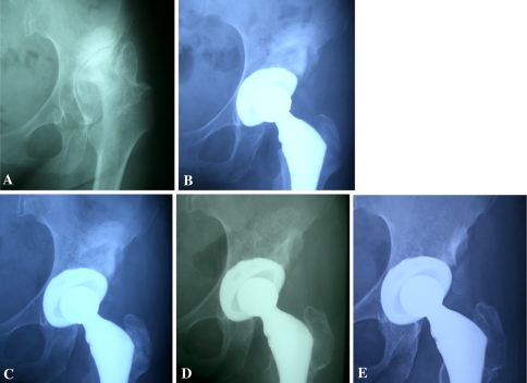
Fig. 6A–E.
The radiographs illustrate the case of a 51-year-old woman with Crowe Type II hip dysplasia. (A) A preoperative radiograph shows acetabular dysplasia. (B) A radiograph taken 3 months postoperatively shows bridging trabeculation. (C) A radiograph taken 12 months after THA shows graft remodeling. (D) A radiograph taken 1.6 years postoperatively shows trabecular reorientation. (E) On the last visit, graft bone volume was unchanged and the interface of the host bone was obscure.

