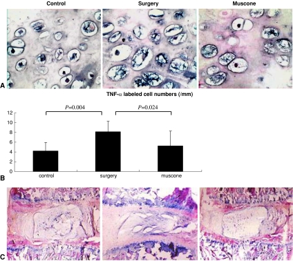
Fig. 7A–C.
(A) The number of TNF-α-positive cells was increased in the surgery group compared with controls but was close to normal in the muscone group (Stain, in situ hybridization staining; original magnification, ×400). (B) Mean values are shown for the number of TNF-α-positive cells in three groups (values shown as means [bars] + SDs [error bars]). (C) In the surgery group, the laminar structures of the annulus fibrosus were disorganized, and the nucleus pulposus was fibrotic and fissured. Muscone treatment almost completely reversed the IVD degeneration caused by surgery (Stain, hematoxylin and eosin; original magnification, ×100).

