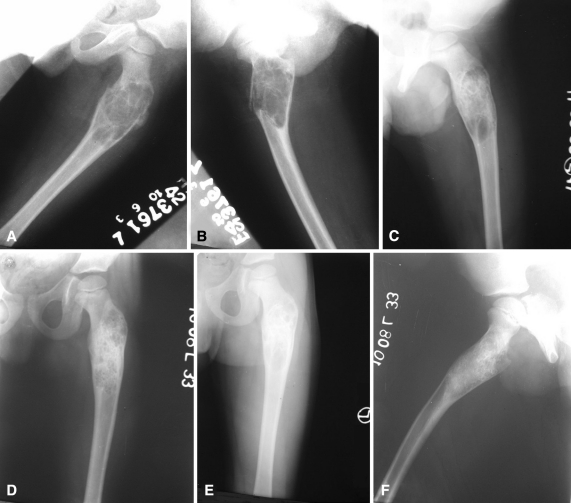
Fig. 1A–F.
(A) AP and (B) lateral radiographs show an ABC affecting the proximal femur of the left side in an 8-year-old boy with pathologic fracture. (C) An AP radiograph taken after two injections of polidocanol shows an unopacified loculation in the lower part of the lesion. (D) A radiograph was obtained at completion of treatment after three injections. (E) AP and (F) lateral radiographs show the lesion at 2 years with remodeling of the lesion.

