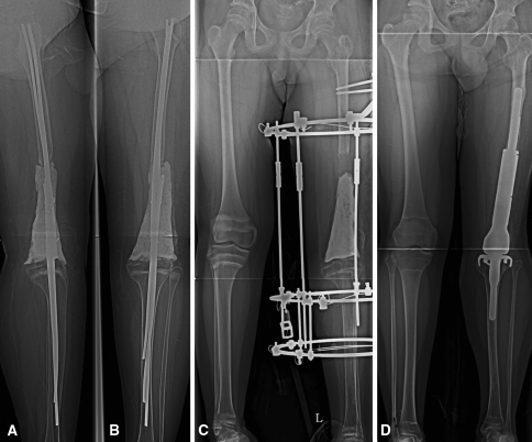
Fig. 1A–D.
Patient 33 was a 12-year-old boy with a diagnosis of metaphyseal osteosarcoma. (A) The lateral and (B) AP radiographs were taken after TRA using multiple Ender nails and bone cement. (C) This AP radiograph was taken after the soft tissue lengthening procedure was finished. The gap between bone cement and the distal end of the femur reveals the amount of length gained. The cement spacer was inserted to maintain room for subsequent implantation. (D) This AP radiograph shows successful conversion to an adult-type implant.

