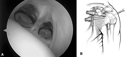
Fig. 2A–B.
(A) An arthroscopic view shows placement of two cannulae, with one at the apex of the rotator cuff interval and a smaller-diameter one inferior and medial just above the subscapularis. (B) A diagram illustrates the two cannulae within the rotator cuff interval. The inferior cannula is above the subscapularis tendon. Reprinted with permission and ©2007 Wolters Kluwer Health from Spencer EE Jr. Partial-thickness articular surface rotator cuff tears: an all-inside technique. Tech Shoulder Elbow Surg. 2007;8:180–184.

