


Figure 2.
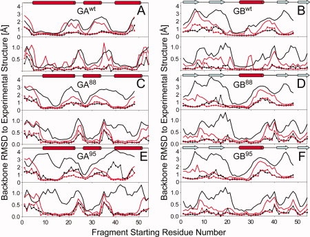
Quality of Rosetta/CS-Rosetta fragments used as input for deriving GA and GB models, shown as plots of the lowest (lines with dots) and average (bold lines) backbone coordinate rmsd's (N, Cα, and C′) between any given segment in the experimental structure and 200 nine-residue (upper panel)/three-residue (lower panel) fragments, as a function of starting position of the query segment. Results from the standard Rosetta fragment selection method are plotted in black, whereas those selected using the standard MFR method with chemical shifts are displayed in red. (A) GAwt; (B) GBwt; (C) GA88; (D) GB88; (E) GA95; and (F) GB95. Note that for nine-residue fragments, the last residue starting number in the 56-residue protein is 48, whereas for three-residue fragments, the last starting position is 54.