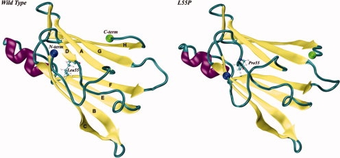
Figure 1.

Schematic representation of the X-ray crystal structures of wild type (on the left) and L55P (on the right) subunits of human transthyretin (TTR; PDB entries 1TTA36 and 5TTR,52 respectively). Residue 55, located in β-strand D, is represented in balls-and-sticks. The N- and C-termini are represented by two spheres. [Color figure can be viewed in the online issue, which is available at www.interscience.wiley.com.]
