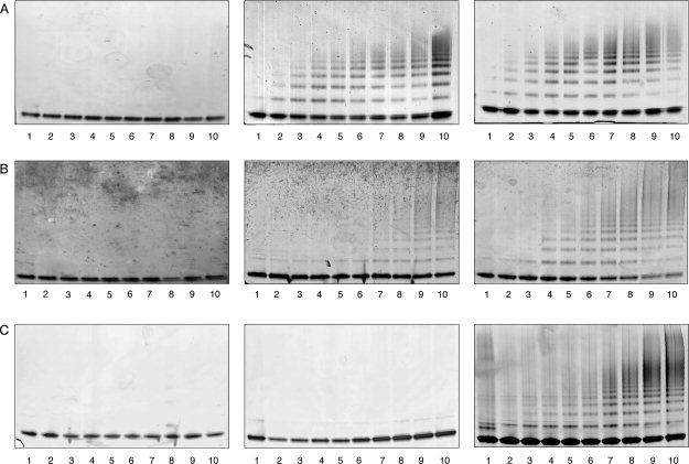
Figure 5.

Neuroserpin mutants were incubated at 45°C and 0.4 mg/mL and aliquots taken over time were analyzed by 3-12% w/v gradient nondenaturing PAGE. All lanes contained 1.2 μg of protein. A: Left: Polymerization of His338Gln neuroserpin at pH 6.0. Middle: Polymerization of His338Gln neuroserpin at pH 7.0. Right: Polymerization of His338Gln neuroserpin at pH 8.0. B: Left: Polymerization of His119Gln neuroserpin at pH 6.0. Middle: Polymerization of His119Gln neuroserpin at pH 7.0. Right: Polymerization of His119Gln neuroserpin at pH 8.0. C: Left: Polymerization of His138Gln neuroserpin at pH 6.0. Middle: Polymerization of His138Gln neuroserpin at pH 7.0. Right: Polymerization of His138Gln neuroserpin at pH 8.0. Lanes 1-10 correspond to 0, 5, 10, 15, 20, 30, 45, 60, 120, and 180 min.
