Figure 3.
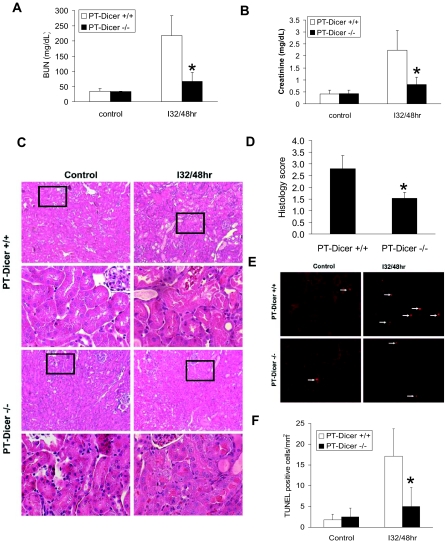
Resistance of PT-Dicer−/− mice to renal IRI. Male PT-Dicer+/+ and PT-Dicer−/− mice of 8 to 10 weeks of age are subjected to sham operation (Control) or 32 minutes of bilateral renal ischemia followed by 48 hours of reperfusion (I32/48h). (A and B) Blood samples are collected to measure BUN (A) and serum creatinine (B). Data are means ± SD. (C) Renal tissues are collected for hematoxylin and eosin staining to examine histology. Histologic images of both low and high magnifications. (D) Ischemia reperfusion–induced tubular damage in PT-Dicer+/+ and PT-Dicer−/− mice is evaluated and scored by histology. Data are means ± SD. (E) Renal cortical and outer medulla tissues are also collected for TUNEL assay of apoptosis. (F) Representative images of TUNEL assay. TUNEL-positive cells are quantified by cell counting in comparable regions of the tissues. Data are means ± SD. n ≥ 4. *Significantly different from PT-Dicer+/+ group.

