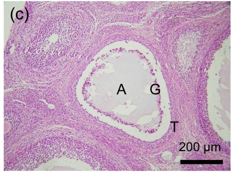
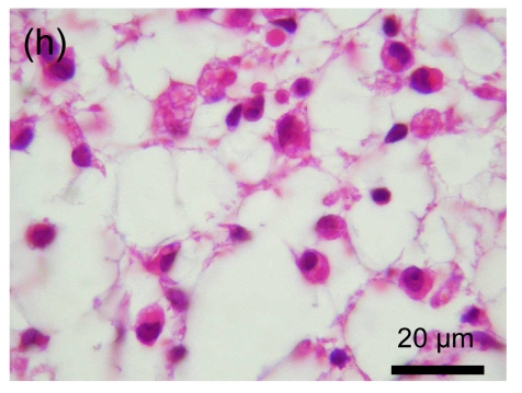
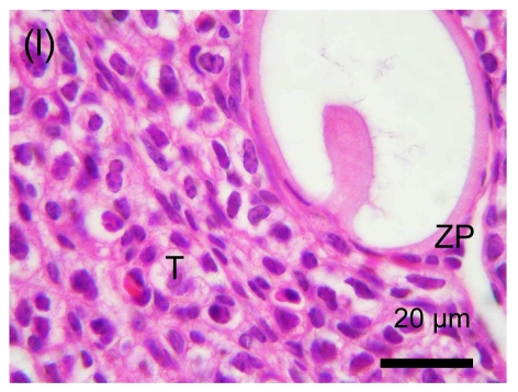
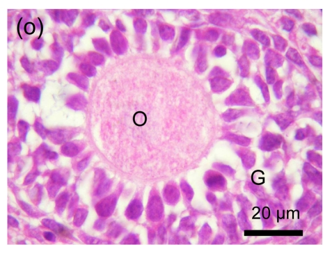
Fig. 1.
Morphologic observation of follicular atresia
(a) & (b) Stage I: granulosa layer became loose and the pyknotic cells appeared; (c), (d) & (e) Stage II: granulosa cells were eliminated massively and floated in the antrum; (f), (g) & (h) Early phase of Stage III: atrophic follicular cavity was occupied by fibroblast-like cells with slender shapes (g) and round shapes (h); (i) & (j) Late phase of Stage III: at first, only a small quantity of differentiated cells appeared around the zona pellucida (i), however, it increased greatly and formed a new tissue subsequently (j); (k) & (l) Stage IV: follicle volume became smaller and smaller; (m) & (n) Abnormal manner of follicular atresia: in some small antral follicles (m), the cumulus granulosa cells were eliminated first and the granulosa layers were relatively intact (n); (o) & (p) Preantral follicular atresia: there was no massive elimination of cells (o), preantral follicles degenerated gradually (p). O: oocyte; G: granulosa layer; T: theca layer; A: follicular antrum; C: Call-Exner body; ZP: zona pellucida


