Figure 1.
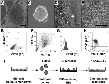
Differentiation of hES cells to hematopoietic progenitors. (A) hES cells were removed from mouse embryonic fibroblasts by treating with collagenase IV and cocultured for 10 days with a monolayer of OP9 in α-minimum essential medium containing 15% fetal calf serum. (B) FACS of a single-cell suspension after 12 days of hES cell coculture with OP9 cells; staining for CD34 and CD43. (C) EBs. hES cells were removed from mouse embryonic fibroblasts by treating with dispase and cultured in EB-differentiating medium for 4 days. (D) Phase-contrast image of adherent EBs differentiated in IL-6, IL-3, SCF, and Fms-like tyrosine kinase 3 ligand for 3 weeks with hematopoietic progenitors releasing to medium (small arrows indicate progenitors; and large arrow, differentiated EBs). (E) Phase-contrast image of homogeneous population of hematopoietic progenitors collected from medium 4 weeks after EB differentiation. (F) Forward-side scatter plot of hematopoietic progenitors. (G) CD34 surface expression on hematopoietic progenitors. Histogram shows antibody staining (in dark) relative to isotype-matched control (transparent). (H) CD45 and CD43 surface expression. Microscopic figures (A,C-E) were captured using an Olympus IX81 inverted fluorescence microscope with 4× (A,C,D) and 10× (E) objectives with a Hamamatsu ORCA RC camera, operated by Velocity software (PerkinElmer Life and Analytical Sciences). Bars in insets represent 100 μm. Data shown are representative from at least 3 experiments. (I) Schema showing coculture-free differentiation of human mast cells from hES cells.

