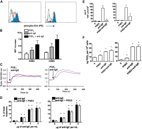
Figure 4.
Activation of ESMCs by cross-linking of FcϵRI and PGE2. (A) Phosphorylation of p42/44 ERK assayed by FACS using a phosphospecific antibody that recognizes phosphorylation of T202 and Y204. ESMCs (left panel) or CBMCs (right panel) were activated by FcϵRI cross-linking or by PGE2 activation (black represents unactivated; green, PGE2; red, anti-IgE; and blue, PGE2 and anti-IgE). (B) Quantitative analysis (n = 6) of T202/Y204 phosphorylation in ESMCs stimulated by PGE2 (white bar), FcϵRI (hatched bar), and FcϵRI and PGE2 together (dark bar). (C) Intracellular calcium release in ESMCs (left panel) or CBMCs (right panel) and activated by FcϵRI cross-linking 3 μg/mL anti-IgE antibody (blue) or 1μM PGE2 (red). Point of stimulation is shown by arrow. Calcium mobilization is expressed as the ratio of fluorescence of Fura-2 measured at 340 and 380 nm. Typical results from at least 2 experiments are shown. (D) Histamine release in ESMCs (left panel) or CBMCs (right panel) preincubated with hIgE and cross-linked with various concentrations of anti-IgE antibody alone or together with 10−6M PGE2. Histamine release is presented as a percentage of total histamine assayed by lysis of cells by Triton X-100 (n = 3). *Significant difference from 0 μg/mL anti-IgE (P < .05). (E) Release of TNF-α was evaluated in ESMCs (left panel) and CBMCs (right panel) preincubated with hIgE and cross-linked with 3 μg/mL anti-IgE antibody alone or together with 10−6M PGE2 (n = 3). (F) PGD2 release from ESMCs (left panel) or CBMCs (right panel) measured using PGD2 methoxime enzyme immunoassay kit (n = 3). *Significantly different from untreated cells (P < .05). Results shown in right panels in panels C and E were obtained under identical conditions to those in the left panels but on different days.

