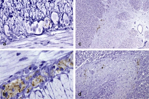
Figure 2.
(a) Trace PrPd deposits in a single ganglion cell of the myenteric plexus of a goat that was positive in 7 of 10 LRS tissues and also in brain (score 1.7). (b) Widespread PrPd accumulation in ganglion and satellite cells of the ENS of a clinically-affected goat (9/10 LRS positive tissues and 20.0 score in brain). Mild PrPd deposition in the intermedio-lateral column (c) and substantia gelationosa (d) of the TSC of a goat that was positive in 7 of 10 LRS tissues and in brain (score 7.0). IHC with Barr224 PrP antibody and haematoxylin counterstaining (magnifications: a, d ×20; b ×40; c ×10). (For a color version of this figure, please consult www.vetres.org.)

