
Figure 1.
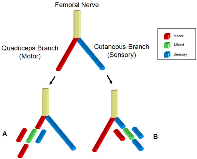
Rodent femoral nerve model used to study modality specific regeneration. Below the femoral bifur cation, either the motor branch to the quadriceps (A) or the cutaneous sensory branch (B) was cut and reconstructed with a motor (red), mixed (green), or sensory (blue) nerve graft harvested from the femoral motor branch, peroneal nerve or femoral sensory branch respectively. Grafts from each group were then harvested at 5, and 7 weeks post transplantation and evaluated using histomorphometry. For the 6 week time point motor grafts or sensory grafts were placed in the transected motor branch of the femoral nerve.