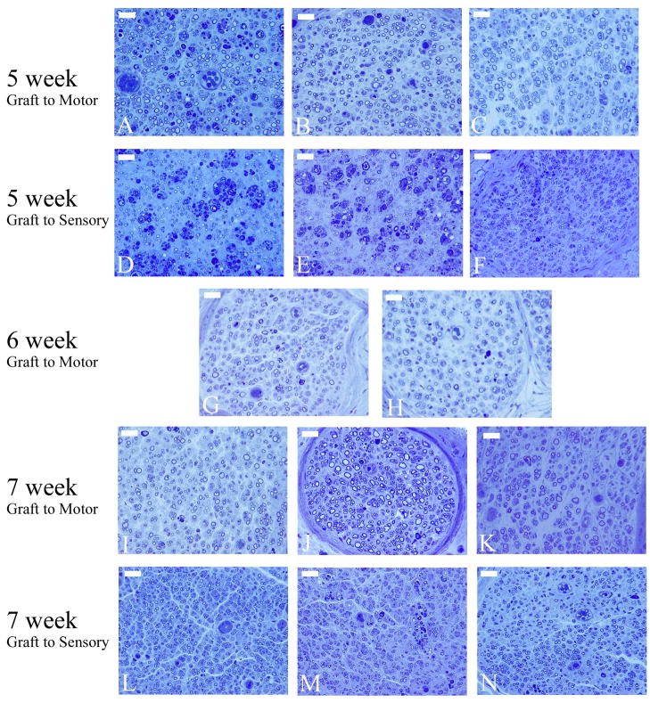
Figure 3.
Representative toluidine blue histological sections used for histomorphometric analysis from each experimental group at each time point. The first two rows of images are mid-graft photomicrographs of motor (A&D), mixed (B&E), and sensory(C&F) grafts bridging the transected femoral motor (A–C) and transected femoral sensory branch (D–F) at the 5 week time point. The third row of images is photomicrographs of the distal stump of the transected femoral motor branch with either a motor graft (G) or sensory graft (H) 6 weeks after grafting. The final two rows of images display photomicrographs from the distal stump of the transected femoral motor (I–K) and femoral sensory (L–N) branches treated with either a motor (I&L), mixed (J&M), or sensory graft (K&N) 7 weeks after grafting. (scale bar = 5 μm)

