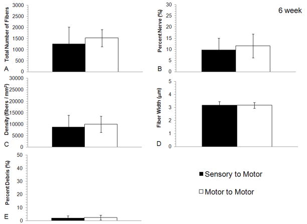
Figure 5.
Histomorphometric comparisons of nerve regeneration in the distal stump of the transected femoral motor branch using either sensory or motor graft modalities after 6 weeks. No differences were seen in the quantification of A) total number of fibers, B) percent nerve, C) fiber density, D) fiber width or E) percent debris between either of the experimental groups regardless of modality match or mismatch (bars are averages, error bars are standard deviation, Motor to Motor n = 10, Sensory to Motor n = 10).

