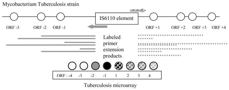
Figure 1.
Schematic description of the biological procedure. We attempt to discern the location of an IS6110 element with straightforward application of microarray technology. Primers specific for the 3′ (right arrow) and 5′ (left arrow) end of the insertion sequence are applied to the genome. Primer extension generates labeled DNA fragments. Upon application of these fragments to the microarray spots representing ORFs closest to the IS6110 element appear intense (black), while spots further from the IS6110 element appear dim (light gray). In the experiment the 5′ and 3′ fragments are labeled with two different dyes so that spots −4 to −1 will be labeled with one dye (solid) and spots 1 to 4 will be labeled with a different dye (dashed).

