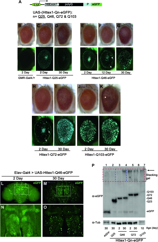
Figure 1.—
Age- and polyQ-length-dependent formation of aggregates in Drosophila. (A) Structure of the Htt exon1 (Httex1) constructs used for the studies. eGFP-tagged Httex1 with different lengths of glutamine tract (polyQ) followed by its proline-rich region (P) are under the control of the UAS elements (Brand and Perrimon 1993). (B–P) PolyQ-length- and age-dependent formation of aggregates in Drosophila. (B–K) Httex1-Qn-eGFP were expressed in the eye using the eye-specific GMR-Gal4 driver. (Top) The general eye morphology of adults of different ages. (Bottom) Images of the same eyes under green fluorescent light, with their age and genotype indicated below. Genotypes: (B) GMR-Gal4/+ and (C–K) GMR-Gal4/+; UAS-Httex1-Qn-eGFP/+. (B) Control of GMR-Gal4 driver alone at day 2. Note that there is no visible eGFP signal in this eye (bottom) in the absence of the UAS-eGFP reporter. (C and D) Adult eyes with Httex1-Q25-eGFP expression. Q25 is found mainly in a diffuse cytoplasmic pattern at both day 2 (C) and day 30 (D). Note that the center bright spots in both eyes and in E are not aggregates but rather an optical phenomenon similar to the “deep pseudopupil,” arising from the superimposition of evenly diffuse GFP signals emitted from the underlying regularly arranged ommatidia (Franceschini 1972). (E–G) Adult eyes with Httex1-Q46-eGFP expression. At day 2 (E, left eye), Httex1-Q46 is found mainly in a diffuse cytoplasmic pattern. As the fly ages, sporadic aggregates gradually accumulate (F, white arrows) and become prominent by day 30 (G). (H and I) Adult eyes with Httex1-Q72-eGFP expression. At day 2 (H), Q72 is found mainly in a diffuse cytoplasmic pattern but sporadic aggregates are already visible (H, white arrows). Aggregates become prominent by day 30 (I). (J and K) Adult eyes with Httex1-Q103-eGFP expression. Aggregates already become prominent at day 2 (J) and persist at day 30 (K). Note also that there is a mild loss of pigment at the posterior of this eye at day 30 (K, white arrowhead), indicating the degeneration of underlying eye tissues at this stage. In all images, the eyes are oriented as dorsal side is up and anterior is to the right. (L–O) Confocal images of aggregates formation in the brain by the Httex1-Q46-eGFP, which was expressed in the CNS using the pan-neuronal Elav-Gal4 driver. Genotypes: Elav-Gal4/+; UAS-Httex1-Q46-eGFP/+. The protein was distributed evenly, and no obvious aggregates were found in the brain at the younger age (L and N, day 2), whereas numerous prominent aggregates were present at day 30 (M and O). (N and O) Higher magnification view of regions (the olfactory bulb and the α- and β-lobes of mushroom body) highlighted in L and M, respectively. Note that the images in the younger brain (L and N) were exposed for a longer time to maximize the detection for aggregates. (P) Confirmation by Western blot of the development of SDS-insoluble aggregates in Httex1-Qn-eGFP flies. Whole-protein extracts from adult fly heads were probed with anti-eGFP antibody. (Bottom) Ages and the type of the expressed protein in the examined flies. Large protein complexes that were retained in the stacking gel, as highlighted, were found in samples from Httex1-Q72, Q103 (lanes 5–7) and aged Q46 flies (lane 4, 30 days old), but were absent in young Q46 flies (lane 3, 2 days old) and other control flies (lane 1, flies expressing eGFP alone; lane 2, Httex1-Q23-eGFP; both were 30 days old).

