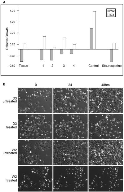
Figure 3.
(A) Change in relative W2 and D3 cell viability by MTT assay 48 h after addition of whole tissue extract, 1, 2, 3, and 4 (1 12, 2 29, 3 12, 4 11 μM). Staurosporine (0.1 μM), a known apoptosis inducer, and untreated cells were used as positive and negative controls. Values for the compounds are an average of five wells, with a standard deviation of less than 15% of the mean. (B) Compound 1 is a specific inducer of apoptosis in mammalian cells. The apoptosis-competent W2 cells and the apoptosis -resistant D3 cells were cultured in the presence or absence of compound 1 (12 μM) under normal physiological conditions (DMEM with 10% FBS, 5% CO2 at 37 °C), and the cell viability was monitored by time-lapse microscopy. D3 cells were viable in the presence of compound 1 beyond 48 h, whereas the apoptosis-competent W2 cells showed massive apoptosis induction and significant loss of viability by 24 h of treatment.

