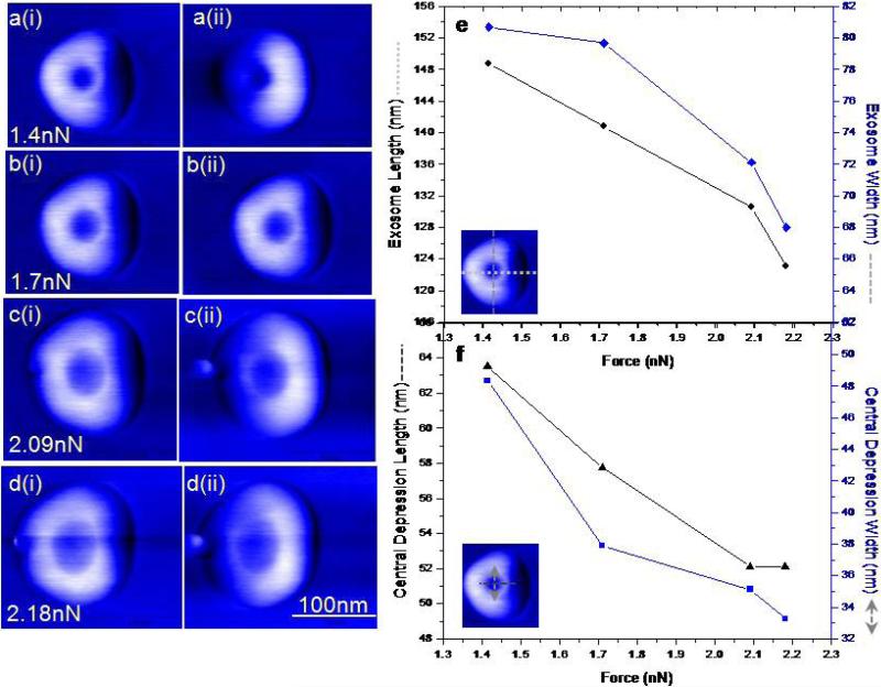
Figure 2. Mechanical deformation of single exosomes during increased imaging force under PM-AFM.
a-d, Consecutive phase images of same exosome showing vesicle size as a function of applied force imaged under forward (i) and backward(ii) scan direction for each force setpoint. Increasing imaging forces (a to d) cause the overall lateral dimensions to increase and the central depression occupies more of the apparent structure. Blebbing of exosomes at high forces(c and d) a bleb or pinch observed on exosome surfaces suggest structural perturbation. Forward tip movement possibly results in backward folding of the bleb on the exosome structure whereas during backward scan the bleb is extended farther away from the exosome displayed as a protrusion. Bleb may result from a combination of mechanical stress well as inherent structural configuration of the exosomes. e, Cross sectional analysis of exosome structure under varying forces. f, Corresponding changes in the size of the central depression. The depth of the depression, changes from 1.1nm (at1.41 nN) to 1.7nm (at 2.18 nN).

