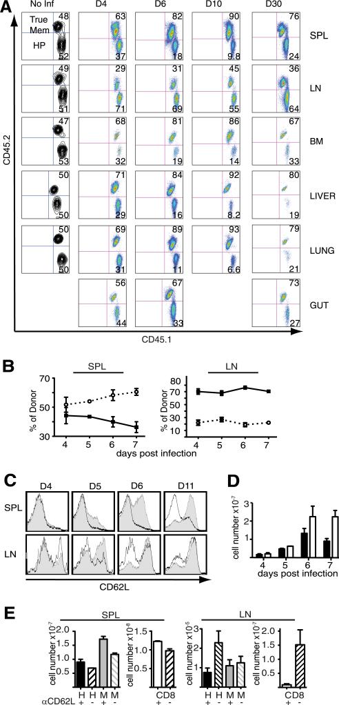
FIGURE 2.
Defect in HP-memory cell accumulation is not due to peripheral localization. The ratios of co-transferred memory subsets were monitored in the indicated tissues after Lm.OVA infection. A, Relative accumulation of the donor (CD45.1+ CD8+) subsets in the spleen (SPL), lymph nodes (LN), bone marrow (BM), liver, lung, and gut following infection. Memory subsets in the spleen and lymph nodes after infection: (B) percent of each donor memory subset (HP, solid line; true-memory, dotted line), (C) CD62L expression on co-transferred memory subsets (HP, gray; true-memory, white). D, Average total cell numbers from pooled spleen and all lymph nodes (HP, black; true-memory, white). E, Total cell numbers for donor HP- or true-memory cells from the spleen (left) and all recovered lymph nodes (right) following treatment with anti-CD62L or PBS (similar results obtained with isotype control). Host naive CD8+ cell numbers indicated in separate graphs (right). Representative of >3 experiments (n = 3). Error bars indicate SD. Mem, memory; Inf, infection; d, day; H, HP; M, true-memory.

