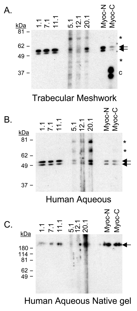
Figure 3. Myocilin detected in human trabecular meshwork tissue and aqueous humor.
Myocilin monoclonal antibodies identified myocilin in (A) human trabecular meshwork tissue, (B) denatured and reduced human aqueous humor, and (C) native human aqueous humor (nondenatured and nonreduced). All antibodies detected myocilin doublet at 53–57 kDa (arrows) in denatured and reduced gels while the N-terminal antibodies showed greater specificity for myocilin in native gels, identifying myocilin above 180 kDa marker (arrow). Asterisks indicate additional myocilin related bands recognized by C-terminal antibodies 5.1, 12.1, and 20.1. Lowercase C indicates C-terminal fragment. [Note: Protein marker in (C) is denatured and reduced therefore reflects accurate size].

