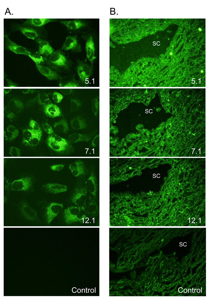
Figure 4. Immunohistochemistry of myocilin monoclonal antibodies.
(A) Myocilin was localized perinuclear with an endoplasmic reticulum/Golgi Apparatus distribution. Representative images from monoclonal antibodies 5.1, 7.1, and 12.1 are shown. All six myocilin monoclonal antibodies showed a similar distribution. 400× magnification. (B) Representative images of myocilin monoclonal antibodies 5.1, 7.1, and 12.1 showing myocilin associated with trabecular cells throughout the meshwork and with Schlemm’s canal cells in paraffin-embedded tissue. 600× magnification.

