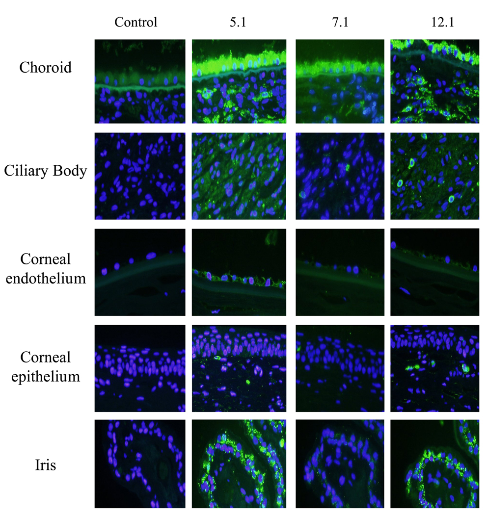
Figure 5. Recognition of myocilin in various tissues of the human eye.
Images of choroid, ciliary body, corneal endothelium, corneal epithelium and iris show identification of myocilin with selective myocilin monoclonal antibodies. From left to right: Control (secondary antibody alone), 5.1 (middle left column), 7.1(middle right column) and 12.1 (right column). Each column represents cross-sections of a single eye. Images were photographed at 40× magnification.

