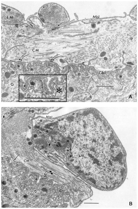
Fig. 4.
The circular muscle (CM) and longitudinal muscle (LM) interface between the gut (G) outer surface is revealed by ultrastructural analysis (A and B). Muscle basal lamina (Mbl) is relatively thin and single-layered, while gut basal lamina (Gbl) is relatively thick and multilayered (arrow) revealing characteristic vertical crosshatch marks (asterisk; inset). Muscle cells present a smooth contour, while the basal aspect of the enterocytes is highly convoluted and deeply folded (asterisk). The Mbl and Gbl are closely juxtaposed, but this approximation is not confluent down into folds (asterisk). Cell junctions between muscle cells (curved arrow) and between muscle cells and enterocytes (double curved arrows) are noted. Scale bar = 0.5 μm.

