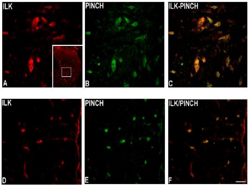
Fig. 3.

Immunoreactivity of ILK in lumbar spinal cord. ILK is present in dorsal horn gray matter (A) and in white matter (D). Similarly, PINCH immunoreactivity is present in dorsal horn gray matter (B) and in white matter (E). Note the coexpression of ILK and PINCH in both gray matter (C) and white matter (F). Bar = 40μm. Inset in A: low-power view of the dorsal horn immunostained for ILK, with the approximate location of higher-power images delimited by the white square.
