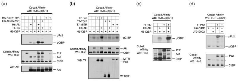
Fig. 2. Akt1 phosphorylates CtBP1 and Pc2.
(a) COS1 cells were transfected with H6-CtBP1, Flag-Pc2, H6-Akt1, H6-Akt(S473D), or HA-Akt(K179A) expression constructs as indicated. Cells were lysed in 6M guanidine-HCl, proteins purified on cobalt agarose and western blotted with an Akt1 substrate [RxRxxp(S/T)] antibody. Lower panels: 20% of the pulldown was analyzed by western blot using H6 and a portion of the lysate with an Akt1 antibody. (b) COS1 cells were transfected with the indicated expression constructs and analyzed as in panel a. Cobalt affinity purified proteins were analyzed with phospho-substrate and His6 antibodies [upper panels] and part of the lysate was analyzed by direct western blot with a T7 antibody [below]. (c) 293T cells were transfected with H6-CtBP1, H6-Akt1, and Flag-Pc2 expression constructs as indicated, and analyzed as for panel a. (d) 293T cells were transfected with H6-CtBP1 and Flag-Pc2 expression constructs and were left untreated or treated with 50μm LY294002 for 1 hour, and analyzed as for panel a.

