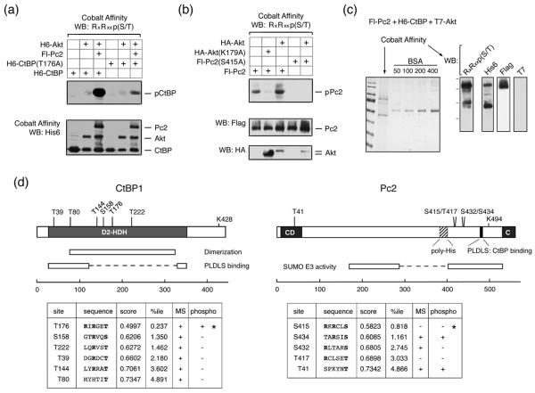
Fig. 5. Identification of phosphorylation sites in CtBP1 and Pc2.
(a) COS1 cells were transfected with H6-CtBP1, H6-CtBP(T176A), Flag-Pc2, and H6-Akt1 expression constructs as indicated and phosphorylation was analyzed by western blot with an Akt phospho-substrate antibody. (b) COS1 cells were transfected with Fl-Pc2 or Fl-Pc2(S415A), together with a control vector, HA-Akt1, or HA-Akt(K179A) expression constructs, and analyzed by western blot as in A. (c) A sample used for MS analysis is shown. Pc2 and CtBP1 were purified from transiently transfected COS1 cells expressing both proteins together with T7-Akt1. A portion of the purified proteins was analyzed by Coomassie blue staining (left), or by western blot with the indicated antibodies (right). (d) CtBP1 and Pc2 are shown schematically, together with the positions of the closest matches to consensus Akt phosphorylation sites. The sequences of consensus Akt phosphorylation sites are shown below, together with the score and percentile as predicted by Scansite. A + in the MS column indicates that peptides spanning the site were identified in the MS analysis. A + in the phospho column indicates that phosphorylation was detected at that site by MS. * indicates the two sites which were identified by western blotting with the phospho-substrate antibody.

