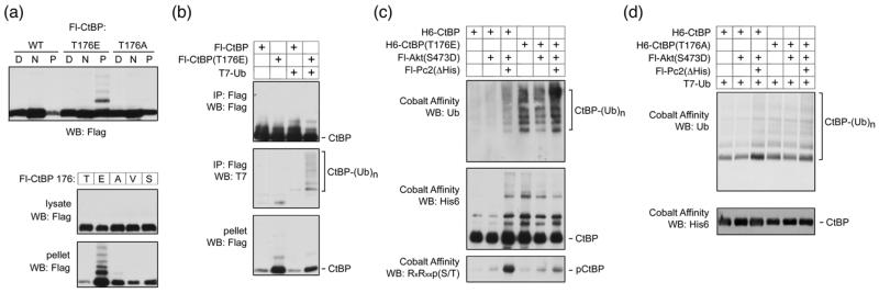
Fig. 9. Ubiquitylation of CtBP1.
(a) COS1 cells were transfected with Flag tagged wild type or mutant CtBP1 as indicated and partitioned into digitonin soluble [D], NP-40 soluble [N], and pellet [P] fractions, which were analyzed by Flag western blot. Lower panels show NP-40 lysate and pellet fractions from COS1 cells expressing Flag-CtBP1, in which residue 176 was either wild type [T], or altered to one of the other indicated amino acids, were analyzed by western blot. (b) COS1 cells were transfected with Flag tagged CtBP1 or the T176E mutant, together with a T7-tagged ubiquitin expression construct, as indicated. Flag immunoprecipitates, or the pellet fraction were analyzed by Flag and T7 western blot as indicated. (c) COS1 cells were transfected with the indicated expression constructs, lysed in guanidine HCl and His-tagged CtBP1 was purified by metal affinity [note the Pc2 construct used here lacks the poly-histidine stretch, so does not bind cobalt agarose]. Bound CtBP1 was analyzed by western blot with antibodies for endogenously expressed ubiquitin, H6-CtBP1 and phosphorylation at T176. (d) COS1 cells were transfected with the indicated expression constructs, lysed in guanidine HCl and CtBP was isolated on cobalt agarose. Bound His-tagged CtBP was detected with a His6 antibody (lower panel) and ubiquitylated CtBP was detected with a T7 antibody (upper panel).

