Figure 2.

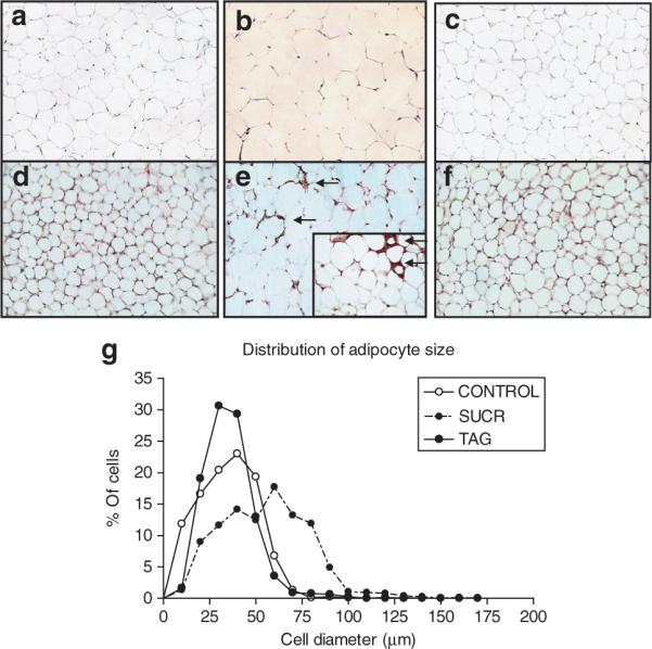
(a–c) Adipocyte diameter and (d–f) macrophage immunostaining are increased in retroperitoneal adipose tissue sections from SUCR-fed compared to TAG and CONTROL LDLr−/− mice. Representative adipose sections from (a) CONTROL, (b) SUCR, and (c) TAG-fed mice, demonstrating larger adipocytes in SUCR compared to TAG and CONTROL mice. F4/80 macrophage immunostaining was not evident in sections from (d) CONTROL or (f) TAG-fed mice (all images are ×20 magnification). In contrast, adipose tissue macrophages were observed in sections from SUCR-fed mice (e; arrows denote positively stained cells; inset is ×40). (g) Distribution of adipocyte size. LDLr−/−, low-density lipoprotein receptor deficient.
