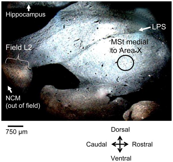
Figure 2.
Darkfield rostroventral section showing the circular template (0.75 mm in diameter, black circle outline in figure) used to sample a control region of medial striatum (MSt) on 10 sections distributed approximately 200 μm-986 μm lateral from the midline. The template was placed ventral to the pallio-subpallialis lamina (LPS) and caudal to the area where Area X would emerge in more lateral sections. Field L is seen as a light band in the caudal region of the brain. NCM is caudal to Field L.

