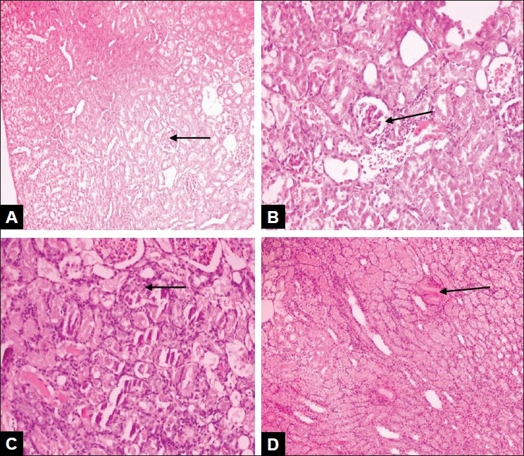
Fig. 1.

Photomicrographs of rat kidneys from different groups (A): Kidney tissue of control animal with normal glomeruli with an intact bowman's capsule and proximal convoluted capsule and stained with Haematoxylin and Eosin at magnification 40X. (B): Kidney tissue of animal treated with gentamicin with glomerular congestion, inflammatory cells, necrosis and tubular casts and stained with Haematoxylin and Eosin, at magnification 200X. (C): Kidney tissue of leaves extract-treated animals with blood vessel congestion, interstitial edema and tubular casts and stained with Haematoxylin and Eosin, at magnification 800X. (D): Kidney tissue of unripe pods extract-treated animals with normal arrangement of cells and stained with Haematoxylin and Eosin, magnification 100X.
