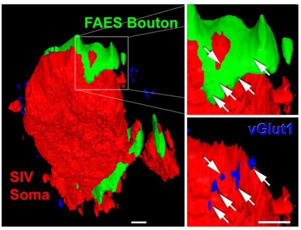
Fig. 2.
A FAES bouton in contact with SIV interneuron co-localizes vesicular glutamate transporter 1, indicative of a functional synapse. The left panel shows a PV-positive interneuron soma (red; appears flat because it was on the cutting plane of the section) in somatosensory area SIV. However, around the perimeter of this interneuron two contacts from auditory FAES axons (green) and numerous vGlut1-positive (blue) points are visible. The right panel shows a digital enlargement of the boxed FAES bouton (green)–SIV interneuron (red) anatomical contact. The white arrows indicate the precise position of vGlut1-immunoreactive points that are visible when the FAES axon is rendered transparent, as depicted in the lower right panel (vGlut1-positive points are blue at arrow tips). The presence of vGlut1 at this axon-interneuron contact is indicative of a functional, glutamatergic synapse. Average voxel dimension ~200 nm, scale bars 1 μm

