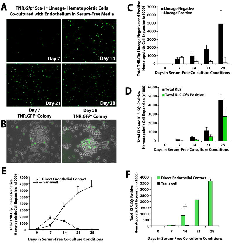
Figure 3. Notch signaling is activated in hematopoietic cells co-cultured with E4ORF1+ ECs and sKitL.
A) One thousand freshly isolated KLS hematopoietic cells from the BM of TNR.Gfp mice were co-cultured with E4ORF1+ ECs and sKitL. The degree of Notch signaling in the culture increased over time as indicated by enhanced Gfp expression (TNR.Gfp+ cells) in the expanding hematopoietic cells. B) Hematopoietic colonies that attach to ECs increase Notch signaling over time. C) Quantification of total number of TNR.Gfp+ Lin− versus Lin+ hematopoietic cell expansion. TNR.Gfp+ hematopoietic cells cultured in the absence of endothelium died by three weeks in culture. D) Phenotypic expression comparing the expansion of total KLS versus TNR.Gfp+ KLS cells expanded in the presence of E4ORF1+ ECs and sKitL. E,F) TNR.Gfp hematopoietic cells cultured in direct cellular contact with E4ORF1+ EC + sKitL or placed on the upper chamber of the transwells, physically separated from E4ORF1+ ECs. Lack of cellular contact of the TNR.Gfp hematopoietic cells with the E4ORF1+ EC monolayers, even in the presence of sKitL, results in a severe impairment of survival and diminished proliferation of the Lin− TNR.Gfp hematopoietic cells (E) and impaired expansion of TNR.Gfp+ KLS cells (F). By contrast, TNR.Gfp hematopoietic cells that were cultured in direct cellular contact with E4ORF1+ ECs and sKitL underwent significant expansion over a 28 day period, generating large numbers of Lin− TNR.Gfp (E) and TNR.Gfp+ KLS cells (F). Error bars represent SD. (*p<0.05, n = 6)

