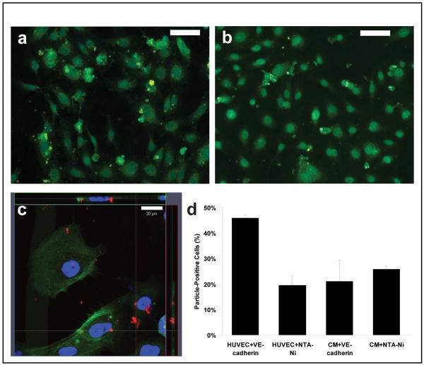
Figure 6. Microparticles can be targeted to HUVEC though VE-Cadherin.
(A) Fluorescence micrograph of HUVECs incubated with VE-Cadherin loaded microparticles. Microparticle cores were loaded with coumarin-6 for tracking. (scale bar 100 μm) (B) Representative fluorescence micrograph of HUVEC incubated with Ni-NTA microparticles. (scale bar 100 μm) (C) Laser scanning confocal micrograph of HUVEC with VE-Cadherin loaded microparticles. Orthogonal projection of z-stack suggests that microparticles are not internalized. (scale bar 20 μm) (D) Percentage of cells positive for at least one microparticle. Negative controls (HUVEC+Ni-NTA, NCM+VE-Cadherin, NCM+Ni-NTA) ranged from 19-26% of cells positive for one microparticle. By contrast, HUVECs incubated with VE-Cadherin loaded microparticles had 45.7% of cells labeled by microparticles, which represents a 2.4-fold increase compared to HUVECs treated with non-targeted microparticles. (data shown: mean ± SEM)

