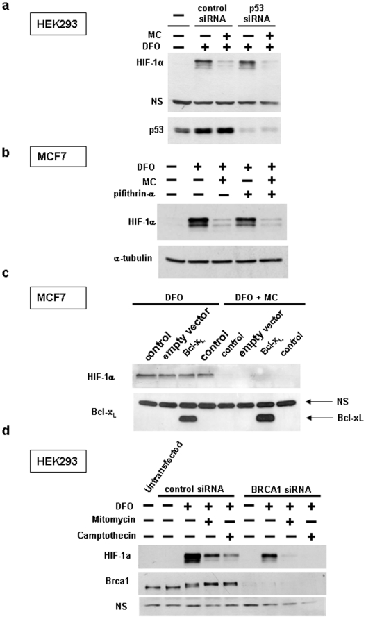
Figure 5. Inhibition of HIF-1α by mitomycin C is independent of p53 and DNA damage-induced apoptosis.
(a) HEK293 cells were transfected with negative control or p53 siRNA for three days, followed by treatement with 200 µM desferrioxamine and 10 µg/ml mitomycin C for 10 hours and Western blotting of cell lysates with the indicated antibodies. (b,c) MCF7 cells were treated with 200 µM desferrioxamine and 10 µg/ml mitomycin C for 14 hours as indicated, followed by Western blotting of cell lysates. In (b) pifithrin-α (25 µM) was included, as indicated. In (c) MCF7 cells were retrovirally transduced with Bcl-xL or empty vector, as indicated. (d) Brca1 was knocked down using siRNA for three days, as described under Materials and Methods, followed by drug treatment (200 µM desferrioxamine, 10 µg/ml mitomycin, 2 µM camptothecin), as indicated.

