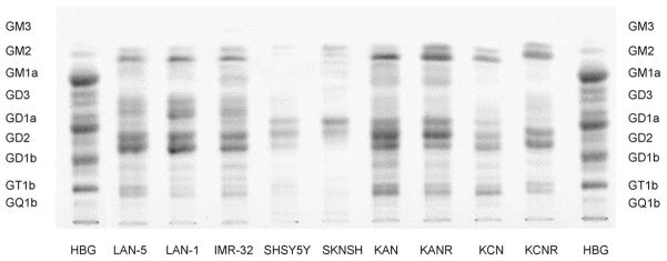
Fig. 2.
Ganglioside expression in nine human NB cell lines (LAN-1, LAN-5, IMR-32, SK-N-SH, SHSY-5Y, SMS-KCN, SMS-KCNR, SMS-KAN, SMS-KANR). Human brain gangliosides (HBG) were employed as a standard. Most tumor ganglioside bands appear as doublets due to ceramide structural heterogeneity. The lower GD1a band and the upper GD2 band overlap.

