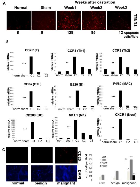
Figure 1. Androgen ablation induces tumor inflammatory infiltration.
Six week old FVB males (n=10) were inoculated with myc-CaP cells. When tumors reached 1000 mm3, mice were left untreated, castrated or sham operated. Tumors were collected when indicated for analysis. A. Paraffin-embedded tumor sections were TUNEL stained to determine apoptotic cell frequency (results are averages, n=3). B. Total RNA was isolated from tumor samples and expression of indicated cell marker mRNAs was quantitated and normalized to that of cyclophilin A (norm= normal, C1,2,3= mice analyzed 1,2,3 weeks after castration, sham= sham-operated). Results are averages ± s.d. (n=10). C. Frozen human prostate sections (normal tissue [n=3], prostatic hyperplasia [n=3], and malignant CaP with Gleason scores 6-8 [n=10]) were stained with CD4, CD8 and CD20 antibodies and DAPI and analyzed by immunofluorescent microscopy. The histogram denotes average frequencies of indicated cell types (n=3 per sample). P values were determined and are depicted as insignificant (ns), significant (*), very significant (**) or highly significant (***).

